Dr. de Clermont-TonnerrePlastic Surgeon

Paris 16 & Pontoise (95)

Melanoma Surgery

Excision & Reconstruction — Paris 16 & Pontoise

Melanoma is the most serious skin cancer but, when diagnosed early, it is almost always cured by surgery. Dr. de Clermont-Tonnerre performs excision per HAS guidelines with immediate aesthetic reconstruction, in Paris 16 and Pontoise.

Warning sign: Any mole that changes (size, shape, colour, bleeding) should be assessed promptly. Do not delay in booking an appointment.

Book an Appointment

Screening

The ABCDE Rule

A

Asymmetry

The two halves of the mole do not match

B

Border

Irregular, scalloped or poorly defined edges

C

Colour

Multiple shades within the same lesion (brown, black, red, blue)

D

Diameter

Larger than 6 mm (the diameter of a pencil eraser)

E

Evolution

Any recent change: size, shape, colour or bleeding

Surgical Management

01

Urgent Consultation

Clinical and dermoscopic examination, lesion assessment. If suspicious, diagnostic excision is scheduled with relative urgency.

02

Diagnostic Excision

Removal of the lesion with narrow margins (2 mm). Histopathological result in 5 to 7 days confirming the diagnosis and measuring tumour thickness (Breslow index).

03

Wide Re-excision + Sentinel Node

Wide excision with recommended margins (1 to 2 cm per Breslow). Sentinel lymph node biopsy under general anaesthesia if Breslow > 1 mm.

04

Reconstruction & Follow-up

Immediate reconstruction by closure, flap or skin graft. Multidisciplinary follow-up plan established (dermatologist, oncologist if required).

Practical Information

Anaesthesia
Local (diagnostic excision) / General (re-excision + sentinel node)
Hospitalisation
Day surgery to 1 night
Time off work
5 to 15 days depending on procedure
Reimbursement
Covered by French national health insurance
Urgency
Prompt consultation recommended

Clinical Examples

Recognising a Melanoma

These images illustrate the ABCDE criteria and typical features of melanocytic lesions to monitor, for educational purposes only. Any suspicious mole should be assessed by a doctor.
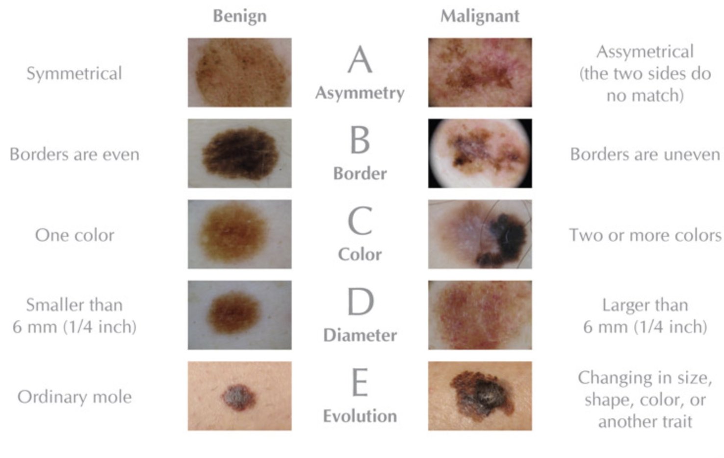
ABCDE rule melanoma — comparison benign and malignant naevus

These images are presented for educational purposes. If in doubt about a lesion, consult a doctor promptly.

Frequently Asked Questions

Is melanoma dangerous?+
Melanoma is the most serious skin cancer due to its metastatic potential. However, when diagnosed at an early stage (in situ or stage I), the prognosis is excellent: five-year survival exceeds 90% for stage I. This is why any suspicious mole should be assessed promptly without delay.
How do I recognise a suspicious melanoma? The ABCDE rule+
The ABCDE rule helps identify suspicious lesions: A (Asymmetry — the two halves do not match), B (Border — irregular or scalloped edges), C (Colour — heterogeneous colour with multiple shades in the same lesion), D (Diameter — greater than 6 mm), E (Evolution — any recent change in size, shape, colour or bleeding). The presence of one or more of these criteria warrants an urgent consultation.
What is the surgical technique for melanoma?+
Excision is performed with safety margins defined by HAS guidelines according to the Breslow index: 0.5 cm for in situ melanomas, 1 cm for melanomas less than 1 mm thick, and 2 cm for melanomas more than 1 mm thick. Immediate reconstruction of the defect is carried out in the same operative session by direct closure, flap or skin graft.
What is a sentinel lymph node?+
For melanomas with a Breslow thickness exceeding 0.8 mm, sentinel lymph node biopsy (the first lymph node draining the tumour) is recommended per HAS guidelines. It is performed under general anaesthesia at the same time as the wide excision. A positive node (invaded by tumour cells) directs towards additional systemic treatment (immunotherapy or targeted therapy).
Is melanoma surgery covered by health insurance?+
Yes, melanoma surgery (diagnostic excision, wide re-excision, sentinel lymph node biopsy, reconstruction) is fully covered by French national health insurance. For thick or metastatic melanoma, long-term condition (ALD 30) status ensures 100% reimbursement of the entire care pathway.
Is follow-up necessary after melanoma excision?+
Yes, lifelong dermatological surveillance is essential after melanoma excision, regardless of tumour thickness. It includes annual (or twice-yearly for stages II–IV) dermatological consultations, imaging according to stage, and regular dermoscopy of remaining naevi. Dr. de Clermont-Tonnerre coordinates this follow-up with the referring dermatologist and oncologist.
Is sun protection mandatory after melanoma?+
Yes, strict and rigorous photoprotection is essential for life after a melanoma diagnosis. This includes avoiding sun exposure during peak hours (11am–4pm), wearing protective clothing and a hat, and systematically applying high-factor sunscreen (SPF 50+) to all exposed areas. UV tanning beds are strictly contraindicated.
Can a person develop more than one melanoma during their lifetime?+
Yes, a history of melanoma is one of the main risk factors for developing a second melanoma. This risk is estimated at 5 to 10% over a lifetime. This is why annual dermoscopic surveillance of all naevi is essential. Fair-skinned individuals, those with numerous moles or a family history are particularly at risk.
How does melanoma differ from an ordinary mole?+
A benign mole (naevus) typically has a regular shape, well-defined borders, uniform brown colour and a stable size. Melanoma is distinguished by one or more ABCDE criteria: asymmetry, irregular borders, heterogeneous colour with black, red or blue areas, diameter exceeding 6 mm, and above all any recent change. Dermoscopy considerably refines this clinical diagnosis.
What is the timeframe between diagnostic biopsy and wide re-excision?+
The interval between the initial diagnostic excision and wide re-excision (with definitive margins according to Breslow) is ideally 3 to 4 weeks, to allow time for the complete histopathological report. Re-excision is then planned as promptly as possible, generally within 4 to 6 weeks after histological confirmation, to avoid leaving residual tumour tissue.